Warwick researchers show that lasers could reduce heart attack mortalities
Every seven minutes, someone in the UK suffers a heart attack. As our life expectancies (and waist lines) increase, cardiovascular diseases, most of which are associated with fatty plaques in our blood vessels, prose an eminent threat to the global population. They claim the lives of a staggering 17.3 million people per year worldwide, which is almost one third of all deaths. However, innovative auto fluorescence imaging techniques may be key to detecting high risk patients in the future, allowing early preventative treatment. Dr Tara Schiller at WMG, University of Warwick, along with fellow scientists from Monash University and the Baker institute, Melbourne, have recently proposed such techniques. They have found that exciting samples in the near infrared light range reveals that rupture prone plaques show intrinsic auto fluorescence, allowing them to be used as a marker for high risk patients.
Innovative auto fluorescence imaging techniques may be the key to detection of high risk patients in the future, allowing early preventative treatment…
Caused by low density lipoproteins (‘bad’ cholesterol) breaching the inner wall of the artery, an atheroma is a hard, fatty plaque composed mainly of white blood cells and lipids. These plaques can remain asymptomatically in blood vessels for decades, however some more unstable varieties are susceptible to rupturing. If this occurs, it can induce thrombus formation, or blood clotting. This can rapidly slow blood flow or occlude vessels entirely, leading to a myocardial infarction (heart attack) or stroke. High risk plaques can be characterised by the presence of intraplaque haemorrhages, which is essentially the bursting of blood vessels within the plaques themselves.
Previous methods of plaque composition analysis involved identifying intrinsic fluorescence in the violet and blue range (325–475 nm), emitted by components of the lipid core such as elastin, collagen and oxidized low-density lipoproteins. However, low wavelength fluorescence is not exclusive to lipid core associated materials, meaning this process is limited by low specificity and cannot reliably or selectively identify the high-risk plaques.
This new method of intervascular imaging may prove crucial to the pharmaceutical industry for potential use in preclinical models to monitor plaque stabilising drugs…
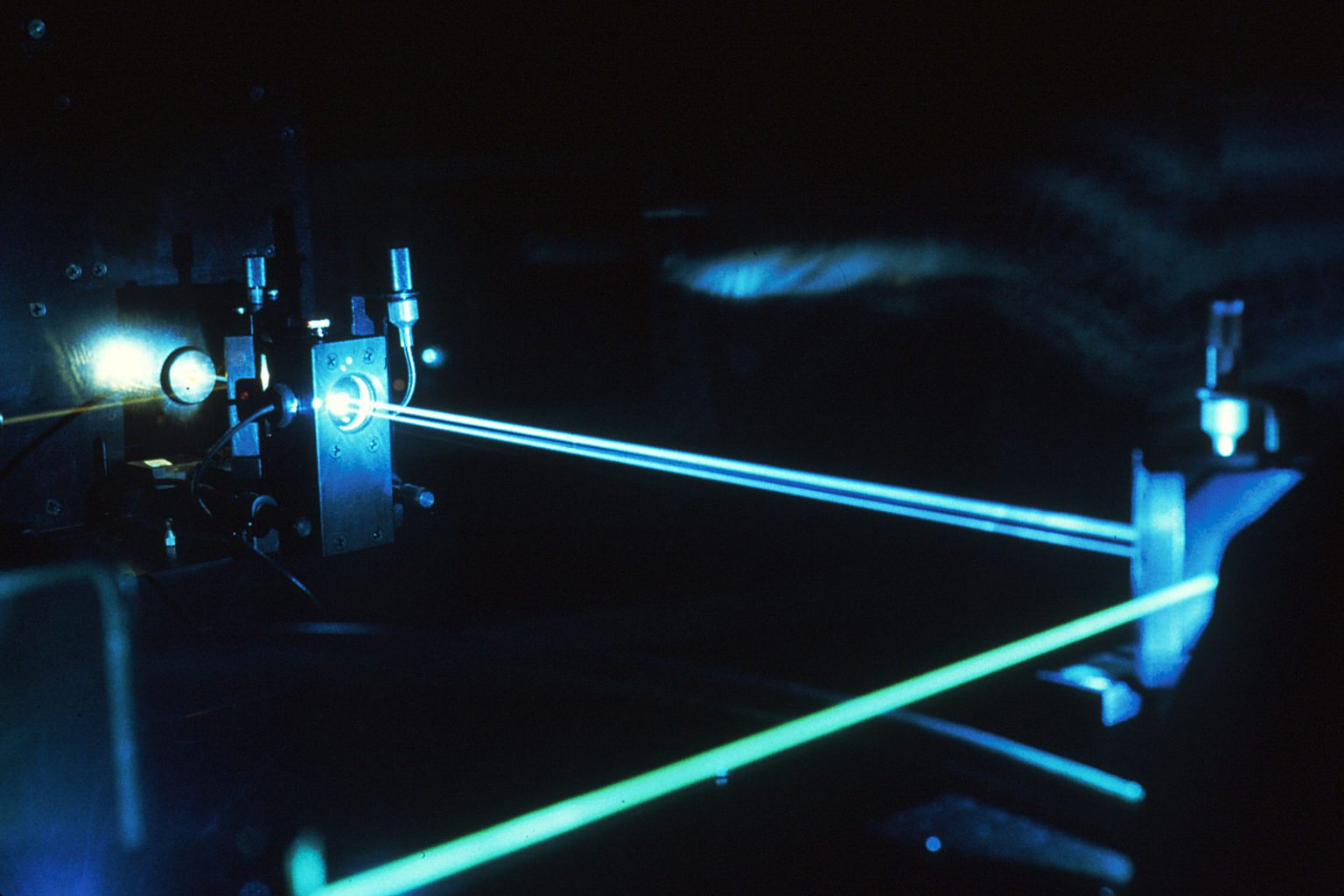
Dr Schiller from the International Institute of Nanocomposites Manufacturing at WMG and colleagues have found that fluorescent measurements in the near infrared range (650–900 nm) show higher tissue penetration, lower photon absorption, and provide the ability to selectively identify deposits that are most vulnerable to rupture. Raman spectroscopy was used to identify the auto fluorescent source as bilirubin, a downstream product of red blood cell degradation. Bilirubin is induced by inflammatory cells in the event of an intraplaque haemorrhage.
In terms of use as a diagnostic tool, near infrared auto fluorescence technology still has to undergo clinical trials before conclusions can be made about its value in atherosclerotic disease prevention and management. However, this new method of intervascular imaging may prove crucial to the pharmaceutical industry for potential use in preclinical models to monitor plaque stabilising drugs. Because of this, it could prove to markedly reduce the morbidity and mortality of cardiovascular disease in the future.
Comments