Warwick research aids understanding of cancer development
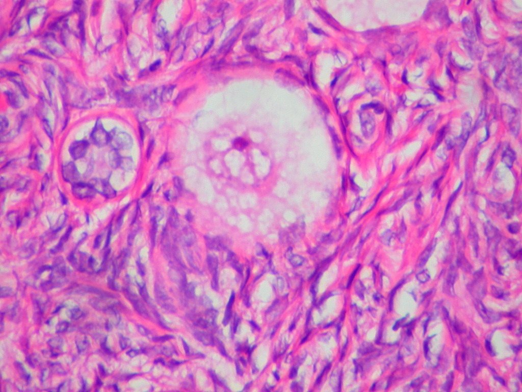
Researchers at the University of Warwick have discovered a cell structure called ‘the mesh’ which could aid scientists’ understanding of the development of cancer cells.
‘The mesh’ helps to hold together cells and has altered the understanding of the cell’s internal scaffolding. Its discovery has been published in the online journal eLife.
The structure was discovered by a team led by Dr Stephen Royle, associate professor and senior Cancer Research UK Fellow at the division of biomedical cell biology at Warwick Medical School.
The discovery of ‘the mesh’ was accidental, with researchers finding it while looking at gaps between microtubules, which are part of the cells’ ‘internal skeleton’.
One of Dr Royle’s PhD students was examining structures in dividing cells using a technique called tomography which is similar to a small-scale version of a hospital CAT scan.
A cell needs to share chromosomes, which carry genetic information, accurately when it divides. If the two new cells end up with the wrong number of chromosomes, a condition that has been linked to a range of tumours in different body organs can develop.
Researchers at the University believe that ‘the mesh’ is needed to give structural support during the division of chromosomes.
It was discovered that one of the proteins that make up the mesh, TACC3, is over-produced in certain cancers. When this situation was mimicked in the lab, the mesh and microtubules were altered and cells had trouble sharing chromosomes during division.
The study received funding and support from Cancer Research UK and North West Cancer Research (NWCR), with NWCR funding the research as part of a collaborative project between the University of Warwick and the University of Liverpool.
Dr Royle said: “As a cell biologist you dream of finding a new structure in cells but it’s so unlikely. Scientists have been looking at cells since the 17th Century and so to find something that no-one has seen before is amazing.”
He added: “We had been looking in 2D and this gave the impression that ‘bridges’ linked microtubules together. This had been known since the 1970s.
“All of a sudden, tilting the fibre in 3D showed us that the bridges were not single struts at all but a web-like structure linking all the microtubules together.”
Dr Emma Smith, senior science communications officer at Cancer Research UK, said: “[The research] might be a crucial insight into why this process becomes faulty in cancer and whether drugs could be developed to stop it from happening.”
Anne Jackson, CEO at NWCR, said: “Dr Royle and Professor Ian Prior at the University of Liverpool have made significant inroads into our understanding of the way in which cancer cells behave, which could potentially better inform future cancer therapies.
“As a charity we fund only the highest standard of research, as evidenced by Dr Royle’s work.”
Comments